

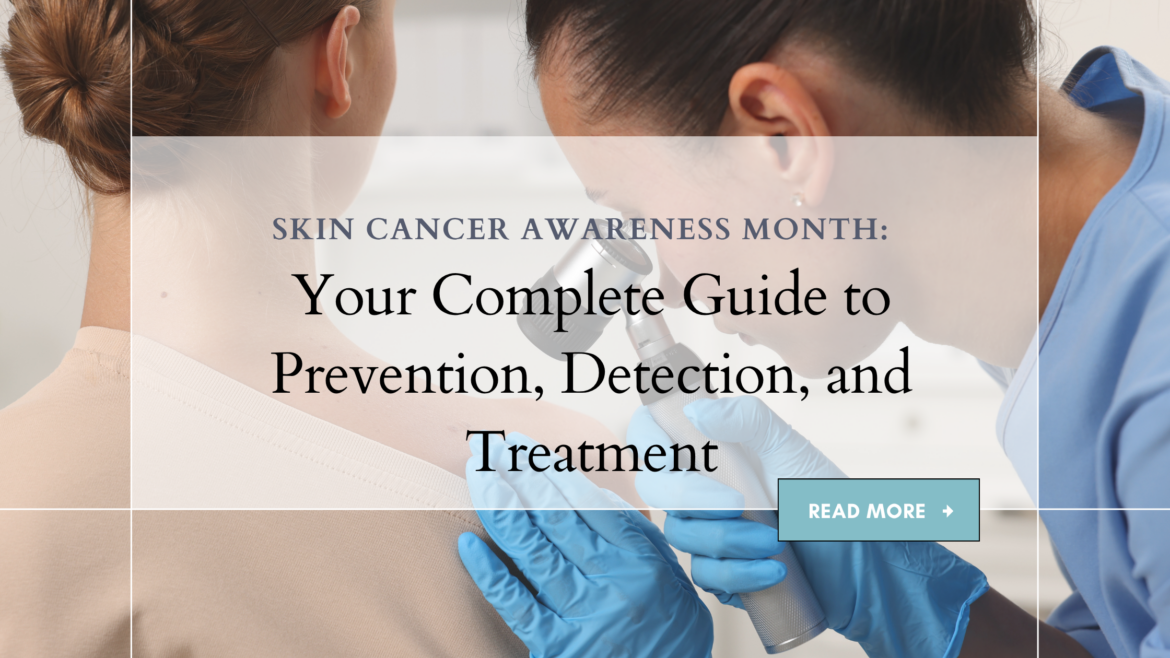
May marks Skin Cancer Awareness Month, a critical time to focus on the most common yet preventable form of cancer in the United States. As a board-certified dermatologist, I witness daily the devastating impact that skin cancer can have on patients and their families, but I also see the remarkable outcomes when these cancers are detected early. With more than 5.4 million cases of skin cancer treated annually in the United States, and one in five Americans developing skin cancer by age 70, education and prevention have never been more important¹.
The statistics are sobering: skin cancer kills more than two people every hour in the United States, yet when caught early, most skin cancers are highly curable². This paradox underscores the critical importance of awareness, prevention, and regular screening. Understanding the different types of skin cancer, recognizing warning signs, and implementing effective prevention strategies can literally save lives.
Understanding the Spectrum of Skin Cancer
Skin cancer encompasses several distinct types, each with unique characteristics, risk factors, and treatment approaches. The three most common types—basal cell carcinoma, squamous cell carcinoma, and melanoma, account for the vast majority of cases, but their behavior and prognosis vary significantly.
Basal Cell Carcinoma (BCC): The most common form of skin cancer, BCC develops in the basal cells of the epidermis. According to the American Academy of Dermatology, BCC accounts for approximately 80% of all skin cancers³. While BCC rarely metastasizes, it can cause significant local tissue destruction if left untreated. These cancers typically appear as pearly or waxy bumps, flat, flesh-colored or brown, scar-like lesions, or bleeding or scabbing sores that heal and recur.
Squamous Cell Carcinoma (SCC): The second most common skin cancer, SCC arises from squamous cells in the outer layer of skin. SCC has a higher metastatic potential than BCC, particularly when it occurs in high-risk locations or in immunocompromised patients⁴. SCC often appears as firm red nodules, flat lesions with scaly or crusted surfaces, or new sores or raised areas on old scars or ulcers.
Melanoma: Though less common than BCC and SCC, melanoma is the deadliest form of skin cancer, responsible for the majority of skin cancer deaths. The American Cancer Society estimates that about 99,780 new cases of melanoma will be diagnosed in 2024⁵. Melanoma develops in melanocytes, the cells that produce melanin, and can occur anywhere on the body, including areas not typically exposed to the sun.
Melanoma Monday: A Critical Awareness Initiative
May 5th marks Melanoma Monday, an annual campaign dedicated to raising awareness about melanoma and promoting early detection. This initiative, supported by dermatologists worldwide, emphasizes that melanoma, while serious, is highly treatable when caught early.
The ABCDE Rule for Melanoma Detection:
The ABCDE criteria provide a systematic approach to evaluating suspicious moles and skin lesions:
- Asymmetry: One half of the mole doesn’t match the other half
- Border: Irregular, scalloped, or poorly defined edges
- Color: Multiple colors or uneven distribution of color
- Diameter: Larger than 6mm (about the size of a pencil eraser)
- Evolving: Any change in size, shape, color, elevation, or symptoms
Research published in the Journal of the American Academy of Dermatology demonstrates that public education about the ABCDE criteria significantly improves early melanoma detection rates⁶. However, it’s important to note that not all melanomas follow these rules, and any new, changing, or concerning lesion should be evaluated by a dermatologist.
Melanoma Statistics and Prognosis:
The five-year survival rate for melanoma varies dramatically based on the stage at diagnosis:
- Localized melanoma (confined to the primary site): 99% five-year survival rate
- Regional melanoma (spread to nearby lymph nodes): 68% five-year survival rate
- Distant melanoma (metastasized to distant organs): 30% five-year survival rate⁷
These statistics underscore the critical importance of early detection through regular self-examinations and professional skin screenings.
Actinic Keratosis: The Precancerous Warning Sign
May 24th is Actinic Keratosis (AK) Global Day, focusing attention on these common precancerous lesions that affect more than 58 million Americans. Actinic keratoses are rough, scaly patches that develop on sun-exposed areas of the skin, particularly the face, scalp, ears, neck, forearms, and back of the hands.
Understanding Actinic Keratosis:
AKs develop as a result of cumulative sun damage over years or decades. The lesions represent areas where the DNA in skin cells has been damaged by ultraviolet radiation, causing abnormal cell growth. While not all AKs progress to cancer, studies show that 5-10% of untreated actinic keratoses may develop into squamous cell carcinoma⁸.
Risk Factors for AK Development:
- Fair skin, light hair, and light eyes
- History of frequent sun exposure or sunburns
- Age over 40 (though younger people can develop AKs)
- Weakened immune system
- Certain genetic conditions
Treatment Options:
Modern dermatology offers numerous effective treatments for actinic keratoses:
- Cryotherapy: Liquid nitrogen freezing is the most common treatment
- Topical medications: Including 5-fluorouracil, imiquimod, and diclofenac
- Photodynamic therapy: Light-activated treatment for multiple lesions
- Chemical peels: For widespread facial AKs
- Laser therapy: For specific lesions or areas
Early treatment of actinic keratoses not only prevents potential progression to skin cancer but also improves the overall appearance and health of sun-damaged skin.
The Critical Role of Professional Skin Screening
Regular professional skin examinations by a board-certified dermatologist are the gold standard for early detection of skin cancer. The American Academy of Dermatology recommends annual skin exams for adults, with more frequent screening for high-risk individuals⁹.
What to Expect During a Professional Skin Exam:
A comprehensive skin examination involves inspection of the entire body, from the scalp to the toes, including areas not typically exposed to sunlight. Dermatologists use dermoscopy, a specialized magnification tool, to examine suspicious lesions in detail. This technique can improve diagnostic accuracy by up to 30% compared to naked-eye examination¹⁰.
High-Risk Individuals Requiring More Frequent Screening:
- Personal history of skin cancer
- Family history of melanoma
- Multiple atypical moles
- Fair skin with a tendency to burn
- History of severe sunburns, especially in childhood
- Immunosuppression
- Occupational sun exposure
The Importance of Baseline Documentation:
Professional skin exams often include photographic documentation of moles and lesions, creating a baseline for future comparison. This “mole mapping” can be invaluable for detecting subtle changes over time that might indicate malignant transformation.
Sun Safety: The Foundation of Prevention
While genetics plays a role in skin cancer risk, most skin cancers are preventable with proper sun protection. The World Health Organization estimates that up to 80% of skin cancers could be prevented by avoiding excessive sun exposure¹¹.
Comprehensive Sun Protection Strategies:
Sunscreen Application:
- Use broad-spectrum sunscreen with SPF 30 or higher
- Apply 1 ounce (2 tablespoons) to cover the entire body
- Reapply every 2 hours and after swimming or sweating
- Don’t forget often-missed areas: ears, lips, feet, and the back of the neck
Protective Clothing:
- Wear tightly woven, long-sleeved shirts and long pants
- Choose wide-brimmed hats that shade face, neck, and ears
- Select sunglasses that block 99-100% of UV rays
- Consider clothing with UPF (Ultraviolet Protection Factor) ratings
Behavioral Modifications:
- Seek shade, especially between 10 AM and 4 PM when UV rays are strongest
- Avoid tanning beds, which increase melanoma risk by 75% when used before age 35¹²
- Be extra cautious around reflective surfaces like water, sand, and snow
- Check the UV Index daily and plan outdoor activities accordingly
Special Considerations: Necrotizing Fasciitis Awareness
May 31st marks Necrotizing Fasciitis Awareness Day, highlighting a rare but serious bacterial infection that can have dermatologic manifestations. While not a skin cancer, necrotizing fasciitis represents a dermatologic emergency that requires immediate recognition and treatment.
Understanding Necrotizing Fasciitis:
Necrotizing fasciitis is a rapidly spreading deep infection of the skin and subcutaneous tissues. The infection can occur following minor trauma, surgical procedures, or even without an obvious entry point. Early recognition is critical, as the infection can progress rapidly and become life-threatening within hours¹³.
Warning Signs:
- Severe pain that seems disproportionate to the appearance of the skin
- Rapidly spreading redness and swelling
- Fever and flu-like symptoms
- Skin that feels hot to the touch
- Changes in skin color to red or purple
- Ulcers, blisters, or black spots on the skin
Risk Factors:
- Diabetes
- Kidney disease
- Compromised immune system
- Chronic heart or lung disease
- Use of anti-inflammatory medications
- Alcohol abuse
While rare, necrotizing fasciitis serves as a reminder that not all serious skin conditions are cancerous, and prompt dermatologic evaluation is essential for any rapidly changing or concerning skin symptoms.
The Role of Technology in Skin Cancer Detection
Advances in technology are revolutionizing the detection and monitoring of skin cancer. Artificial intelligence systems are being developed to assist in melanoma diagnosis, with some achieving accuracy comparable to that of experienced dermatologists¹⁴. Additionally, smartphone apps and teledermatology platforms are making skin cancer screening more accessible, particularly in underserved areas.
Dermoscopy and Digital Monitoring:
Digital dermoscopy allows for high-resolution imaging of skin lesions, enabling precise monitoring of changes over time. This technology is particularly valuable for patients with multiple atypical moles or those at high risk for melanoma.
Confocal Microscopy:
This advanced imaging technique allows dermatologists to examine skin lesions at the cellular level without performing a biopsy, improving diagnostic accuracy and reducing unnecessary procedures.
Prevention Through Education and Lifestyle
Education remains the most powerful tool in skin cancer prevention. Studies consistently show that individuals who understand their risk factors and know how to perform self-examinations are more likely to detect skin cancers early and practice effective sun protection¹⁵.
Self-Examination Guidelines:
- Perform monthly self-examinations in good lighting
- Use mirrors to examine hard-to-see areas
- Have a partner help examine your back and scalp
- Take photos of concerning moles for comparison
- Keep a record of any changes or new lesions
Creating a Sun-Safe Environment:
- Install UV-blocking window film in cars and homes
- Provide shade structures in outdoor recreational areas
- Educate children about sun safety from an early age
- Model good sun protection behaviors
- Make sunscreen application a daily habit, like brushing teeth
The Economic Impact of Skin Cancer
Beyond the human cost, skin cancer represents a significant economic burden. The annual cost of treating skin cancer in the United States exceeds $8 billion, with melanoma treatment costs alone approaching $3 billion annually¹⁶. These figures underscore the importance of prevention and early detection, which are far more cost-effective than treating advanced disease.
Conclusion: Taking Action During Skin Cancer Awareness Month
Skin Cancer Awareness Month serves as a crucial reminder that skin cancer, while common and potentially deadly, is largely preventable and highly treatable when detected early. The key messages are clear: protect your skin from UV radiation, perform regular self-examinations, and maintain a relationship with a board-certified dermatologist for professional screening.
As we observe Melanoma Monday, Actinic Keratosis Global Day, and other awareness initiatives this May, remember that awareness without action is insufficient. Schedule your annual skin exam, update your sun-protection routine, and educate your family and friends on skin cancer prevention.
The fight against skin cancer requires a comprehensive approach involving individual responsibility, professional medical care, and community education. By working together, we can reduce the incidence of skin cancer and save lives through early detection and prevention.
Your skin is your body’s largest organ and your first line of defense against the environment. This Skin Cancer Awareness Month, make a commitment to protect it, monitor it, and seek professional care when needed. The life you save may be your own.
Sources:
- American Academy of Dermatology. “Skin cancer facts & statistics.” Available at: https://www.aad.org/media/stats-skin-cancer
- Skin Cancer Foundation. “Skin cancer facts & statistics.” Available at: https://www.skincancer.org/skin-cancer-information/skin-cancer-facts/
- American Academy of Dermatology. “Basal cell carcinoma: Overview.” Available at: https://www.aad.org/public/diseases/skin-cancer/types/common/bcc
- Karia, P. S., et al. “Cutaneous squamous cell carcinoma: estimated incidence of disease, nodal metastasis, and deaths from disease in the United States, 2012.” Journal of the American Academy of Dermatology, 2013.
- American Cancer Society. “Key Statistics for Melanoma Skin Cancer.” 2024.
- Berwick, M., et al. “The role of the ABCDE criteria in the early detection of melanoma.” Journal of the American Academy of Dermatology, 2015.
- American Cancer Society. “Survival Rates for Melanoma Skin Cancer.” 2024.
- Criscione, V. D., et al. “Actinic keratoses: Natural history and risk of malignant transformation in the Veterans Affairs Topical Tretinoin Chemoprevention Trial.” Cancer, 2009.
- American Academy of Dermatology. “How to examine your skin.” Available at: https://www.aad.org/public/diseases/skin-cancer/find/check-skin
- Kittler, H., et al. “Diagnostic accuracy of dermoscopy.” The Lancet Oncology, 2002.
- World Health Organization. “Ultraviolet radiation and the INTERSUN Programme.” Available at: https://www.who.int/uv/en/
- Boniol, M., et al. “Cutaneous melanoma attributable to sunbed use: systematic review and meta-analysis.” BMJ, 2012.
- Stevens, D. L., & Bryant, A. E. “Necrotizing soft-tissue infections.” New England Journal of Medicine, 2017.
- Esteva, A., et al. “Dermatologist-level classification of skin cancer with deep neural networks.” Nature, 2017.
- Robinson, J. K., et al. “Education on sun protection and skin self-examination.” Dermatologic Clinics, 2006.
- Guy, G. P., et al. “Years of potential life lost and indirect costs of melanoma and non-melanoma skin cancer.” PharmacoEconomics, 2011.